
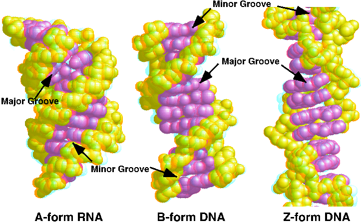
A, B and Z forms, depending upon salt and water concentrations and sequence.
Original Xray studies of DNA done using fiber of bulk DNA (B-DNA) or simple copolymer DNA (A-DNA). The molecules were not aligned in a three-dimensional lattice, like true crystals, so the resultant pattern could not be resolved to atomic level detail. Models were built to fit the available data.
DNA structures are now determined using crystals of synthetic oligonucletides. This has improved the precision of the description of DNA structure, but we will see there is still some ambiguity about the structure of Z-DNA
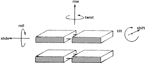
The geometry of the DNA forms can be used to describe the differences seen.
| A | B | Z | |
| Helix sense | Right handed | Right-handed | Left handed |
| Repeating unit | 1 bp | 1bp | 2 bp |
| Rotation/bp | 33.6° | 35.9° | 60°/2 |
| Mean bp/turn | 10.7 | 10.0 | 12 |
| Inclination of bp to axis | +19° | -1.2° | -9° |
| Rise/bp along axis | 2.3Å | 3.32Å | 3.8Å |
| Pitch/turn of helix | 24.6Å | 33.2Å | 45.6Å |
| Mean propeller twist | +18° | +16° | 0° |
| Glycosyl angle | anti | anti | C: anti, G: syn |
| Sugar pucker | C3'-endo | C2'-endo | C: C2'-endo, G: C2'-exo |
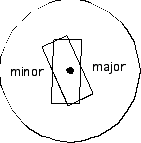
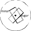
| Diameter | 26Å | 20Å | 18Å |
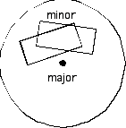
Grooves and stacking of bases:
Major and minor groove definition:
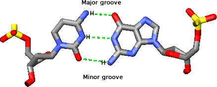
Sugars are on minor groove side of
the base pair
| A | B | Z |
 |
 |
 |
| RNA and
low humidity DNA Bp displaced from axis-> deep major groove |
Hydrated
DNA Bp on axis, both major and minor grooves available |
High salt DNA (Pu-p-Py polymers) bp stick out into major groove. |
Sugar pucker:
Differences --> differences in forms
of helices
| A | B | Z |
 |
 |
 |
| C3'-endo (favored in RNA due to steric problems with 2'OH) | C2'-endo | C: C2'-exo
G: C2'-endo |
| Glycosyl (c) ant | Glycosyl (c) anti | C: Glycosyl
(c) anti G: Glycosyl (c) syn |
Most of variations in conformation
come from variations in d and c.
Can plot them like Ramachandran plots for proteins.
 |
 |
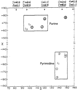 |
| A-form: mostly clustered in one region | B-form: continuum of values, but d related to c. | Z-form: tight clusters of values, but differ for purine and pyrimidine. |
Notes from web tutorial:
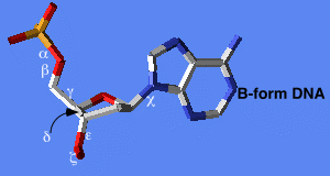
B-form
Now the bases are easier to see. Notice how they are stacked upon each other and are nearly perpendicular to the axis of the double helix. Note also that the backbone forms a smooth, continuous curve.
Zoom in on a few base pairs with
this button. Hydrogen bonds between the bases are shown in white. You are looking
into the major groove. Each base pair stacks on the next similarly, as shown
from this view. A-form DNA also stacks in this way, but compare this with Z-DNA,
which behaves much differently. DNA is usually found in the B form under physiological
conditions. Sometimes kinks are found in the B helix at transcriptional control
regions. These kinks can either be intrinsic to the DNA sequence or caused by
transcription factor binding.
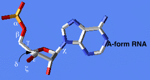
A-form
Notice how they are stacked upon each other but not perpendicular to the axis of the double helix. They are also displaced to the side of the axis. The result is a wide, short helix. Note also that the backbone forms a smooth, continuous curve.
Zoom in on a few base pairs with
this button Hydrogen bonds between the bases are shown in white. You are looking
into the major groove. Each base pair stacks on the next similarly, as shown
from this view. B-DNA also stacks in this way, but compare this with Z-DNA,
which behaves much differently. Essentially all helical RNA is in A form, but
DNA can also be found in A form under certain conditions (particularly in RNA-DNA
hybrids). The 2'-OH of ribose (shown in white in this view) favors the C3'-endo
sugar pucker necessary for A-form geometry. The O2' stericly disfavors the C2'-endo
conformation favored in B-DNA.
Z-form
Notice how the blue bases stack well
on the adjacent blue ones, but not on adjacent red ones, and vice versa. So
it is the dinucleotide unit, rather than mononucleotide that is the repeating
unit of the structure. This explains the need for alternating purines and pyrimidines
to form Z-DNA. You can see the same view without the backbone. Going 5' to 3',
there is good stacking within the GpC dinucleotide, but not between them (CpG).
A top view also illustrates the stacking arrangement. . You can also see this
view without the backbone.