Hammerhead Ribozyme
References:
1. Pley, H.W., Flaherty, K.M. and McKay, D.B. (1994) Three-Dimensional
Structure of a Hammerhead Ribozyme. Nature 372, 68-74.
[Medline Abstract]
2. Scott, W. G., Finch, J. T. and Klug, A. (1995). The Crystal
Structure of an All-RNA Hammerhead Ribozyme: A Proposed Mechanism
for Catalytic Cleavage. Cell. 81, 991-1002. [Medline Abstract]

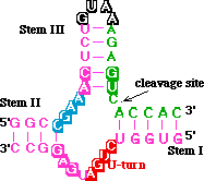
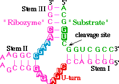
This is the crystal structure of the hammerhead ribozyme from
reference 2. To color the structure as in reference
1, click here <

To see the atoms oriented as in Fig. 3 of reference 1,
click here<
Fig. 4 of reference 1 shows the "domain II"
region of the hammerhead <
>. This region of conserved residues includes two G·A
base pairs, an unusual A-U pair and a standard G-C pair. To see
the h-bonds of the distal half of domain II click here
<>. To see the H-bonds of the proximal
half, click here <>.
You can also see this structure side-by-side
with the first one. There is also a red/blue stereo picture of both structures available.
 Nucleotide
numbering help.
 tRNA
structure.
 Hammerhead
Ribozyme - Main Tutorial (Ref. 1).
 Side-by-side
Comparison of Hammerhead Structures.
 Red/blue
stereo picture of both Structures.
 Back
to intro to DNA-RNA structure.
Comments or Suggestions to:Jim
Nolan at jnolan@tulane.edu


![]() Back
to intro to DNA-RNA structure.
Back
to intro to DNA-RNA structure.