Pathogenesis of Invasive Amebiasis
Click on numbers for explanations
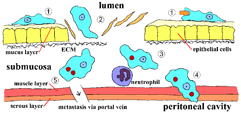
E. histolytica trophozoites colonize the mucosal surface of the large intestine. The trophozoites adhere to the mucus layer and ingest bacteria and cellular debris from the lumen. Adherence is mediated by a protein called the eh-lectin which is expressed on the surface of trophozoites. This non-invasive infection is usually asymptomatic, or exhibits symptoms ranging from mild abdominal discomfort to diarrhea and cramps. [↑Top][↓Bottom]
A breakdown in the mucous barrier can lead to a contact-dependent killing of the epithelial cells. Proteases secreted by the trophozoites may weaken the mucus. Necrotic and apoptotic mechanisms are probably involved in the killing of the host cells. This necrosis of the mucosa will lead to an invasive disease characterized by dysentery (ie, blood and mucus in the feces). In addition, the breakdown of the tissue and extracellular matrix (ECM) implies that proteases are also involved in the pathogenesis. [↑Top][↓Bottom]
The trophozoites will continue to advance laterally and downward into the submucosa producing a 'flask-shaped' ulcer. Necrotic material is found in the center of the ulcer and most of the ameba are at the border between the healthy and damaged tissue. Neutrophils and other immune effector cells are also killed. The ameba are now ingesting host cells instead of bacteria and hematophagous trophozoties can be observed. Ulcers can coalesce and lead to the shedding of patches of mucosa. The severity of the dysentery increases in terms of the number of stools and the amount of mucus and blood. [↑Top][↓Bottom]
The trophozoites can also penetrate the muscle and serous layers leading to intestinal perforations. Perforation of the intestinal wall is a dramatic event that can lead to peritonitis or leakage into the abdominal cavity. Erosion of blood vessels can lead to massive hemorrhage. An inflammatory thickening of the intestinal wall, called an ameboma, or amebic granuloma, can also be formed in response to the ameba. The ameboma presents as a painful palpable mass that can be mistaken for a tumor. [↑Top][↓Bottom]
Trophozoites can also gain access to the circulatory system and be disseminated. The liver is the primary site of extraintestinal amebiasis and hematogenous spread to other organs is rare. Metastasis to the liver involves the portal vein which carries blood from the colon directly to the liver. Dissemination to other tissues most often entails the direct extension of hepatic or colonic lesions. Extraintestinal amebiasis is often characterized by ameba free stools. [↑Top][↓Bottom]
(Note: Print in landscape for best results.)
Use Back Button to return to previously location, or go to:
These pages are developed and maintained by Mark F. Wiser, Tulane University (ã2000). Last update on June 30, 2000.