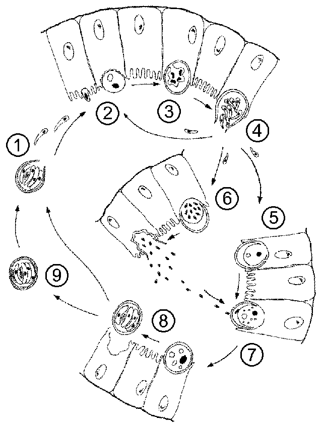
Legend
- The infection is acquired through the ingestion of sporulated oocysts. Motile sporozoites emerge through an opening in the oocyst wall and attach to intestinal epithelial cells. This attachment and subsequent fusion of the microvilli (see below) are probably mediated by the rhoptries and micronemes found at the apical end of the sporozoite. Top
- The sporozoite does not invade the epithelial cell, but induces the fusion of microvilli so that it becomes surrounded by a double membrane of host origin. This location is referred to as being 'extracytoplasmic'. The parasite undergoes a trophic period in which it probably derives nutrients from the host cell via an 'adhesion zone'. Top
- The trophozoite undergoes an asexual replication, called either merogony or schizogony, resulting in the production of 4-8 merozoites. Top
- Mature merozoites are released into the intestinal lumen and will infect new intestinal epithelial cells. Merozoites are morphologically similar to sporozoites and possess apical organelles. The merozoites can either undergo additional rounds of merogony, resulting in the production of more merozoites, or undergo a sexual cycle known as gametogony (or gamogony). Top
- Some of the merozoites will develop into macrogamonts (also called macrogametocytes) which mature into macrogametes. Top
- The microgamont (or microgametocyte) will undergo several rounds of replication producing numerous microgametes which are released into the intestinal lumen.Top
- A microgamete will fuse with a macrogamete (still located in its extracytoplasmic position) and the resulting zygote undergoes sporogony.
Top
- Sporogony involves two rounds of replication resulting in four sporozoites. Fully sporulated oocysts are shed into the intestinal lumen at the completion of sporogony.Top
- The infectious oocysts are excreted in the feces, thus completing the life cycle. An autoinfection in which excystation takes place within the same host may also be possible. It is proposed that the autoinfection is mediated by 'thin-walled' oocysts. Top
|
