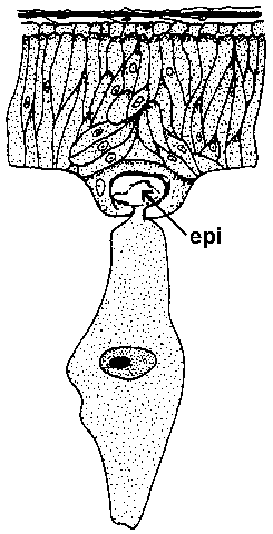
Schematic representaion of the attachment of a gregarine trophozoite to intestinal epithelium. The epimerite (epi) is embedded into the host cell. Modified from Manwell (1961) Introduction to Protozoology.
|
| Schematic representaion of the attachment of a gregarine trophozoite to intestinal epithelium. The epimerite (epi) is embedded into the host cell. Modified from Manwell (1961) Introduction to Protozoology. |
Gregarines are a diverse group of apicomplexan parasites that infect invertebrates--especially arthropods, annelids (segmented worms), and mollusks. The gregarine life cycle typically only consists of gametogony and sporogony and only a few species exhibit merogony. Instead the sporozoites will generally develop into large extracellular trophozoites. In addition, many gregarines do not exhibit intracellular stages. Instead trophozoites attach to the host cell via either a mucron or epimerite (depending on species). These specialized structures are derived from the conoid at the apical end (see Apicomplexa) and function to anchor the parasite to the host (Figure). This attachment to the host cell also functions in feeding in that the cytoplasm of the host is taken up by the attached parasite (i.e., myzocytosis). Gametogony begins when two trophozoites pair, referred to a syzygy, and develop into a gametocyst. Gametes are formed, conjugate, and develop into sporulated oocysts within the gametocyst. The gregarines are thought to be the earliest lineage of apicomplexans.
Analysis of small subunit ribosomal RNA gene sequences (see
Molecular Phylogeny) indicates
that Cryptosporidium is more closely related to the gregarines than
to coccidian (1). In addition, the electron-dense junction observed between
the host cell and Crypotosporidium is morphologically similar to the
attachment site of some gregarines to their host cells (2). However, the morphology
and life cycle is more similar to the coccidia
and thus Cryptosporidium has usually been grouped with the other coccidia.